Kazeti German Shepherds - Exhibitors and breeders of long coat German Shepherds


Before You Do Something Permanent, Know About Growth Plates

growth plates

Before you do something that could impact your puppy’s growth pattern (like an early spay/neuter, or physical activity that’s too vigorous),  you should know when the growth plates close on an average puppy.

Growth plates, also known as the epiphyseal plates or physis, are “zones” of cartilage that exist at the end of bones in both canine and humans as each grows older. They contain rapidly dividing cells that allow bones to become longer until the end of puberty in both humans and canines.  Growth plates gradually thin as hormonal changes approaching puberty signal the growth plates to close, and in most puppies, this is around the age of approximately 18 months old. At that point, the plates “close” because they’ve contributed all they can to the growth of the bones. The growth plate becomes a stable, inactive, part of the bone, but before then, the plates are soft and vulnerable to injury. An injury to the growth plate might not heal properly, nor heal in time for a puppy to grow up straight and strong. Such an injury can result in a misshapen or shortened limb, and that in turn can create an incorrect angle to a joint which can make the puppy more prone to even more injuries when he grows up.

But what about neutering a dog? How that that impact a growth plate?

Part of the responsibility of sex hormones is to regulate growth. When the sex hormones are removed,  growth hormones are missing important regulatory input and the bones continue to grow longer than they ought to.  Growth plates lay down bone as a puppy develops and, as it builds bone, the bone becomes longer and the puppy gets larger and taller. Once maturity is reached, this growth plate turns into bone and the puppy’s full height is reached. Most breeders can spot the difference between an intact dog and a dog neutered too young, and studies have proved it to be true (Salmeri et al, JAVMA 1991).

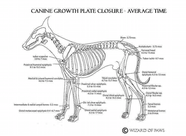
We have the kind permission of Deb Gross to share this fabulous chart,  but we ask that you include the following links if you’re going to share it. Click here to see a larger size.

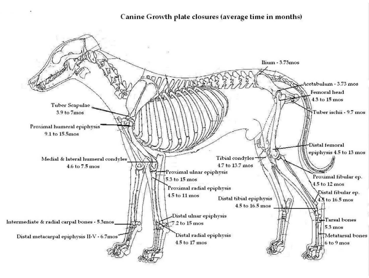
Chart by Deb Gross of Wizard of Paws
Facebook page: https://www.facebook.com/Wizard-of-Paws-95319629966/timeline/
http://www.wizardofpaws.net/


29 thoughts on “Before You Do Something Permanent, Know About Growth Plates”

  1. It was concluded that with respect to skeletal, physical, and behavioral development, the effect of neutering pups at 7 weeks old was similar to that of neutering pups at 7 months old. – A direct quote from Salmeri : so the report does not conclude that which the author of this post professes it to.

    • An interesting comment, Rob. We were able to find what we think is the source of your quote from Dr. Salmeri (https://www.ncbi.nlm.nih.gov/pubmed/2045340) and noted that the same abstract mentioned a delay in growth plate closures in early neuters as compared to dogs who were neutered at an older age which may have resulted in longer bones. This hints at a connection to hip dyplasia.

      In a study of early neuters in Golden Retrievers, early-neutered males had double the occurrence of Hip Dysplasia compared with intact males (Torres de la Riva G, Hart BL, Farver TB, Oberbauer AM, Messam LLM, et al.

      Van Hagen et al (Am J Vet Res, Feb 2005), found that of dogs in their study diagnosed with hip dysplasia, those that were neutered six months prior to the diagnosis were nearly twice as likely to develop hip dysplasia, and in a study by Dannuccia et al (Calcif Tissue Int, 1986), they found that removing the ovaries of Beagles caused increased remodeling of the pelvic bone, which also suggests an increased risk of hip dysplasia with spaying.

      Chris Zinc DVM PhD DACVP said, “…if the femur has achieved its genetically determined normal length at eight months when a dog gets spayed or neutered, but the tibia, which normally stops growing at 12 to 14 months of age continues to grow, then an abnormal angle may develop at the stifle. In addition, with the extra growth, the lower leg below the stifle likely becomes heavier (because it is longer), and may cause increased stresses on the cranial cruciate ligament,” so now we have a connection to cruciate tears. Furthermore, Martin et al, Bone 1987 suspected that sterilization caused a loss of bone mass, and coupled with the fact that neutered females are twice as likely to become obese, the combination can lead to an increased risk of cranial cruciate ligament tear and hip dysplasia. Spayed/neutered dogs are also three times more likely to suffer from patellar luxation (Vidoni et al, Wien Tierartztl Mschr 2005). One has to suspect, then, the possibility that the increase in bone length that results from early-age neutering results in changes in joint conformation, which can lead to hip dysplasia, and other maladies.

      Early spays also hint at a connection to disease. In a study of early neuters in Golden Retrievers, almost 10 percent of early-neutered males were diagnosed with Lymphosarcoma which is three times more than intact males.

      A February 2014 study completed on over 2500 Vizsla dogs found that removing a quarter of the dog’s endocrine system might not be in the dog’s best interest since early spayed females had significantly higher rates of hemangiosarcoma (nine times higher) than intact females. Spayed/neutered dogs were 3.5% more likely to get mast cell cancer, and 4.3 times more likely to suffer lymphoma. The Vizsla study found that the younger the dogs were when spayed/neutered, the younger they were when diagnosed with cancer. (M. Christine Zink, DVM, PhD et al., Evaluation of the risk and age of onset of cancer and behavioral disorders in neutered Vizslas (JAVMA, Vol 244, No. 3, February 1, 2014). We presume that the Dr. Zink mentioned above is the same Dr. Zink who commented on femur length earlier in our comment.

      A Gerald P. Murphy Cancer Foundation study in 2009 found a correlation between the age at which female Rottweilers are spayed and their lifespan. The study compared female Rotties who lived to be 13 or older with a group who lived the expected lifespan of about 9 years. Dr. David J. Waters, a professor in the Department of Veterinary Clinical Sciences at Purdue University said, “”Like women, female dogs in our study had a distinct survival advantage over males. But taking away ovaries during the first [four] years of life completely erased the female survival advantage. We found that female Rottweilers that kept their ovaries for at least [six] years were [four] times more likely to reach exceptional longevity compared to females who had the shortest lifetime ovary exposure.”

      We actually had a chance to attend one of Dr. Waters’ seminars in which he discussed his “Old Gray Muzzle” tour of Rottweilers, and he offered compelling evidence of why neutering isn’t necessarily beneficial to a dog, let alone early neutering.

      This is a lengthy response to your comment which is valid, and we appreciate that you wrote it. However, given the data, early spays and neuters still sound risky.